Sequential size change of F-HUC during Formation of DAS structure on Si(111)
Formation mechanism of the Si(111)7x7 reconstruction studied by scanning tunneling microscopy:
Zipper-like restructuring in the sequential size changes of isolated single faulted-halves
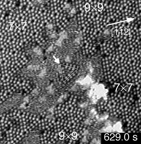
Figure 1. Constant current STM image of the '1x1' region (no atomic-resolution) prepared by quenching to 653 K. An isolated single F-HUC (Faulted-Half Unit Cell) is outlined, being assigned to the 10x10-F. This was taken 629.0 s after the start of measurment.
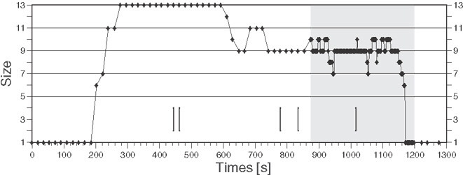
Figure 2. Size changes of the single isolated F-HUC (outlined by a triangle in Fig. 1) as a function of time. The Movie above corresponds to a shaded time-range.